Respiratory and Circulatory Systems
Pharynx and Atrium
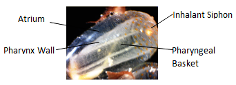
The inhalant siphon opens into a large pharynx, commonly known as the pharyngeal basket. At the junction of the siphon and the pharynx is the mouth, encircled by buccal tentacles (Ruppert et al., 2004). These tentacles protrude into the water current and prevent the entry of large, unwanted particles. The walls of the pharynx are perforated by small ciliated gill slits that allow water to pass through into the surrounding atrium (Ruppert et al., 2004). On the ventral side of the pharynx is the endostyle extending the length of the pharynx. The endostyle divides into two ciliated bands at the junction of the pharynx and inhalant siphon. these bands encircle the pharynx opening (Ruppert et al., 2004).
|
|
E. diaphanis zooid with visible respiratory structures labelled.
|
A water-filled atrium surrounds the pharynx with the exception of the pharynx site of attachment to the body wall (Ruppert et al., 2004). To help support the pharynx cord like mesentries expand across the atrium from the atrial to the pharyngeal walls. The atrium opens to the exterior through the exhalant siphon. The cavity just inside the exhalant siphon is often known as the cloaca; it receives waste from the anus and gametes from the gonoducts (Ruppert et al., 2004).
Respiratory System
Respiratory gas exchange occurs across the surface of the pharyngeal basket, oxygen is taken up from the water current passing through. Due to their low metabolic rate, they have a low oxygen demand with less than 10% of the available oxygen being absorbed (Meglitsch and Schram, 1991) The surface area of the pharyngeal basket is increased for gas exchange by the presence of pharyngeal gill slits and lateral cilia. There are no specialized respiratory organs. Gases are transported to tissues and organs in the plasma.
Circulatory System
Ascidians have a well developed hemal system that includes a circuit through which blood flows, this circuit comprises of heart, vessels and small sinuses. The blood has a rich diversity of hemocytes, but lacks a respiratory pigment (Ruppert et al., 2004).
|
|
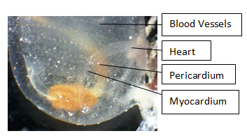
Photograph of an E. diaphanis heart, showing basic features.
|
The heart is a short curved cylinder at the bottom of the digestive loop (Ruppert et al., 2004). It consists of a cylinder with its outer wall folded inwards forming an inner and outer tube. The outer tube is a fluid-filled pericardial cavity and the inner tube surrounds the blood-filled lumen. The wall of the lumen, the myocardium, is formed by a muscular wall of the pericardium folded back upon itself; the outer tube wall is the pericardium (Ruppert et al., 2004). The myocardium contains contractile filaments arranged in two crossed helixes with an angle of 60 degrees between them. The action of these helices causes the heart to contract in a twisting, wringing motion passing from one end of the heart to the other, pushing blood forwards and preventing backflow (Ruppert et al., 2004).
The blood vessels are not lined with endothelia and are simple channels within the connective tissue (Ruppert et al., 2004). There is also no respiratory protein within the blood. It is possible that the large surface area of the pharyngeal basket, the large volume of water pumped and low metabolic activity all make up for the absence of a respiratory protein (Ruppert et al., 2004). The hemocytes found within the blood probably have a variety of other functions, including tunic synthesis, internal defense and defense against predators and competitors (Ruppert et al., 2004).
The hemal circuit is not well established and understood, but the locations of major vessels and the pattern of flow through the pharynx are well known (Ruppert et al., 2004). From the anterior end of the heart, blood enters into a large ventral aorta. The ventral aorta provides the gill capillaries of the pharynx with blood. The capillaries lead to a mid-dorsal vessel, the dorsal aorta. The dorsal aorta carries blood to the gut and viscera. Vessels from these viscera return blood to the heart. At both ends of the heart can be found tunic vessels (Ruppert et al., 2004).
The heart undergoes periodic heartbeat reversal, generally occurring every few minutes. The heart will stop briefly then resume beating in the opposite direction. It is thought that this may be due to the organs and tissues being arranged in series along the blood circuit (Ruppert et al., 2004). Heartbeat reversal would allow those organs at the end of the circulatory circuit to become the first, giving them access to a high concentration of blood-borne nutrients and oxygen. Therefore, it is possible that this reversal is an adaptation designed to average the supply of metabolites to tissues (Ruppert et al., 2004).
The heartbeat is myogenic and is controlled by pacemakers at each end of the heart (Ruppert et al., 2004). These pacemakers alternate in dominance and initiate the opposite directions of beat. Heartbeat reversal may be triggered by pacemaker fatigue or rising back pressure (Ruppert et al., 2004).
|
|
|
Video showing the heartbeat pattern of E. diaphanis. Video taken using a digital microscope, Heron Island, 2013. |
The blood often contains several kinds of hemocytes that can be grouped into three categories: totipotent lymphocytes, which form the connective tissue and give rise to all other types of cells; amebocytes, including phagocytic macrophages; and vacuolated cells, including morula cells and nephrocytes (Ruppert et al., 2004). A peculiarity of ascidian blood is the amoebocyte cells, which accumulte and store excretory waste and the morula cells which accumulate toxins for storage or deposition in the tunic (Pechenik, 2010).
|